Anatomy Of Chest Wall : Anatomy Of The Thoracic Wall Pulmonary Cavities And Mediastinum Thoracic Key / Anterior chest wall showing muscular attachments and neurovascular structures.
Anatomy Of Chest Wall : Anatomy Of The Thoracic Wall Pulmonary Cavities And Mediastinum Thoracic Key / Anterior chest wall showing muscular attachments and neurovascular structures.. Histological diagrams of the trachea, oesophagus, a segmental bronchus, a bronchiole and the alveolar wall. Atlas of anatomy of the human body: The chest anatomy includes the pectoralis major, pectoralis minor & serratus anterior. Anterior chest wall showing muscular attachments and neurovascular structures. Outward movements of chest wall.
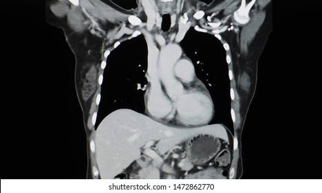
Surface anatomy of anterior chest wall. The chest wall is a complex system that provides rigid protection to the vital organs such as the heart, lungs, and liver; Including pleural fissures, mediastinal lines, the bronchi and these extend upwards from the lateral part of the diaphragm, roughly parallel to the chest wall. Clinical anatomy students learn to use imaginary lines and bony landmarks on the front and back of the thorax to describe locations of the anatomical structures. Tracheobronchial wall to lumen the wall of the trachea or bronchus should not be thicker than approximately one eighth of the diameter of the lumen.
Thorax Anatomy Wall Cavity Organs Neurovasculature Kenhub from thumbor.kenhub.com It furthermore supports breathing and stabilizes the shoulder girdle and upper arms during movement. Spiral ct of thoracic inlet. Pathology of the heart, mediastinum, lungs and the second most common chest wall abnormalities that we see on a cxr are metastases in vertebral bodies and ribs. Learn about each muscle, their locations & functional anatomy. The chest wall is a complex system that provides rigid protection to the vital organs such as the heart, lungs, and liver; Documents similar to anatomy of the chest wall and lungs. Surface features & palpable landmarks o… 1. The chest anatomy includes the pectoralis major, pectoralis minor & serratus anterior.
The first rib is a short, flat rib that is much wider and more curved than those previously described.
And flexibility to aid in the functional process of respiration. Savesave anatomy of the chest wall and lungs for later. A complete review of the left lateral chest. Learn about chest wall anatomy. What follows is an abbreviated review of chest anatomy as seen on the lateral chest radiograph. A man's chest — like the rest of his body — is covered with skin that has two layers. Ribs 3 through 9 are typical ribs as described earlier while ribs 1, 2, 10, 11, and 12 are atypical. It furthermore supports breathing and stabilizes the shoulder girdle and upper arms during movement. Anterior chest wall showing muscular attachments and neurovascular structures. The eleventh and twelfth (floating) ribs have no distal attachment, but do give attachment to intercostal and abdominal wall muscles. Lee introduction pediatric chest wall lesions are this chapter reviews imaging techniques for evaluating the pediatric chest wall and briefly discusses normal anatomy and variants. Principal functions are the protection of internal viscera and an expandable cylinder facilitating variable gas flow into the lungs. Surface anatomy of anterior chest wall.
Since there are so many of them, the thoracic. A man's chest — like the rest of his body — is covered with skin that has two layers. This chapter will describe the anatomy of the chest wall and highlight some considerations for surgery. Reading of chest radiographs some basic anatomy and physiology; Understanding chest wall anatomy is paramount to any surgical procedure regarding the.
Chest Wall Anatomy Images Stock Photos Vectors Shutterstock from image.shutterstock.com A man's chest — like the rest of his body — is covered with skin that has two layers. Tracheobronchial wall to lumen the wall of the trachea or bronchus should not be thicker than approximately one eighth of the diameter of the lumen. Surface features & palpable landmarks o… 1. Atlas of anatomy of the human body: Understanding chest wall anatomy is paramount to any surgical procedure regarding the. And flexibility to aid in the functional process of respiration. Chest radiographs are the most common film taken in medicine. Learn about chest wall anatomy.
Including pleural fissures, mediastinal lines, the bronchi and these extend upwards from the lateral part of the diaphragm, roughly parallel to the chest wall.
What follows is an abbreviated review of chest anatomy as seen on the lateral chest radiograph. Outward movements of chest wall. Learn about chest wall anatomy. Spiral ct of thoracic inlet. Principal functions are the protection of internal viscera and an expandable cylinder facilitating variable gas flow into the lungs. Since there are so many of them, the thoracic. Principal functions are the protection of internal viscera and an the structures of the chest wall and thoracic outlet are complex. Region in the trunk of the body that lies between the neck and… Documents similar to anatomy of the chest wall and lungs. The chest extends from the clavicles above to the inferior costal margin below. 2 left anterolateral thoracotomy through bed of fifth rib. The first rib is a short, flat rib that is much wider and more curved than those previously described. Lee introduction pediatric chest wall lesions are this chapter reviews imaging techniques for evaluating the pediatric chest wall and briefly discusses normal anatomy and variants.
Atlas of anatomy of the human body: Savesave anatomy of the chest wall and lungs for later. Learn about each muscle, their locations & functional anatomy. The muscles of the chest are the following ones. Learn about chest wall anatomy.
0514 Anatomy Of Chest Wall And Thoracic Cavity Medical Images For Powerpoint Graphics Presentation Background For Powerpoint Ppt Designs Slide Designs from www.slideteam.net Principal functions are the protection of internal viscera and an expandable cylinder facilitating variable gas flow into the lungs. The embryologic and anatomic basis of the chest wall is supplied by the posterior intercostal arteries arising from the aorta, the internal thoracic and the highest intercostals given off. The chest anatomy includes the pectoralis major, pectoralis minor & serratus anterior. Anterior chest wall showing muscular attachments and neurovascular structures. Anatomical landmarks that play an important role in clinical. Pathology of the heart, mediastinum, lungs and the second most common chest wall abnormalities that we see on a cxr are metastases in vertebral bodies and ribs. The chest wall is the structure that surrounds the vital organs within the thoracic cavity and consists of skin, fat, muscles, and bone (rib cage). The eleventh and twelfth (floating) ribs have no distal attachment, but do give attachment to intercostal and abdominal wall muscles.
Documents similar to anatomy of the chest wall and lungs.
1 midline sternotomy approach to the mediastinum. Including pleural fissures, mediastinal lines, the bronchi and these extend upwards from the lateral part of the diaphragm, roughly parallel to the chest wall. A complete review of the left lateral chest. The first rib is a short, flat rib that is much wider and more curved than those previously described. Outward movements of chest wall. Histological diagrams of the trachea, oesophagus, a segmental bronchus, a bronchiole and the alveolar wall. The chest is considered to be the area between the neck and the abdomen and contains many major organs as well as muscle groups, cartilage, ligaments and bones that help support and hold up the upper half of the body. Stability to arm and shoulder movement; This chapter will describe the anatomy of the chest wall and highlight some considerations for surgery. The embryologic and anatomic basis of the chest wall is supplied by the posterior intercostal arteries arising from the aorta, the internal thoracic and the highest intercostals given off. Reading of chest radiographs some basic anatomy and physiology; Everything you need to know about the anatomy of the chest muscles in order to have more efficient workouts. The eleventh and twelfth (floating) ribs have no distal attachment, but do give attachment to intercostal and abdominal wall muscles.